Showing posts with label spider anatomy. Show all posts

Thursday, March 05, 2020

Spider with a broken leg

Since we're looking at spiders ...

Here's one I looked at under the microscope.

He's dead; that's why he's sitting still for me.

Zooming in a bit. He's a male he's wearing the boxing-glove pedipalps.

I thought I could identify him by his eye arrangement, but I can't be sure; wrong angle! It doesn't matter, though; what I thought was interesting was the broken leg.

Spider legs are hydraulic. The circulatory system extends through the legs, and the pressure is voluntary. To extend a leg, the spider raises the pressure of the hemolymph (spider blood), which exerts pressure on the joints in his legs, straightening them. To retract the leg, he has muscles.

When the spider dies, the hydraulic pressure drops and the muscles shrink. This is why dead spiders have their legs contracted.

Look at the photos above: the leg in cross-section is basically a tube with a few fibres of muscle, and a wide open space for the hemolymph.

Cross-section diagram. The pale areas on the left drawing are hemolymph space; the darker mid-sections are muscles.*

The legs are connected to the prosoma—the central body of the spider—and are almost completely filled by muscles. Due to the open blood circulatory system, all of the space between the muscles and the exoskeleton—called lacunae—is filled with hemolymph. ... the biological spider uses fluidic expansion of inflatable joint membranes to extend the legs. (From Robotics Open Access Journals *)

* Photo and quote from Landkammer, Stefan et al. “Biomimetic Spider Leg Joints: A Review from Biomechanical Research to Compliant Robotic Actuators.” Robotics 5 (2016): 15. (Common Access)

Oh, and this is why a little spider can take down a huge spider, in spite of her potent fangs: the smaller spider injects her venom at the weaker joints of the legs, and the poison is instantly transported throughout the victim.

~~~~~~~~~~~~~~~~~~~~

Ya que estamos mirando a las arañas, aquí está una que examiné con el microscopio.

Photos: una araña muerta, macho; se puede saber el sexo porque tiene los pedipalpos hinchados. Pensé que podría identificarlo por el arreglo de los ojos, pero no; el ángulo no me deja ver claro. Pero no importa: lo que me interesó fue la vista al interior de esa pata rota.

Las arañas dependen de presión hidráulica para mover sus patas. El sistema circulatorio se extiende hasta el interior de las patas, y el control de la presión es voluntario. Para estirar las patas, la araña aumenta la presión, y eso hace que las articulaciones se enderezen. Para retraer la pata, tiene músculos.

Por eso, cuando una araña se muere, se contraen las patas; ya no hay presión.

Mira las fotos arriba: la pata rota es básicamente un tubo con un poquito de fibra muscular, y un amplio espacio vacío donde antes había hemolinfa (sangre de araña).

La cita y diagrama que sigue son de unos científicos que están estudiando la anatomía de las arañas para aplicaciones en el campo de la robótica.

Otra cosa: esto explica como es que una arañita pequeña puede matar a una araña gigante. La arañita inyecta su veneno por medio de las articulaciones de las patas, que son más débiles, y lejos de los quelíceros de su presa, y el veneno de inmediato se propaga por todo su cuerpo.

Thursday, April 23, 2009

At home in the garden

It was Earth Day. And I spent most of it up to my elbows in the stuff. Earth.

First, I re-potted and trimmed all but one of my houseplants. Then I moved outside, and repotted most of the outside container plants, divided the hostas, transplanted some of the London Pride*, trimmed the evergreens, ripped out a mountain of moss, top-dressed the second half of the garden with manure, and repositioned a wire fence.

A pleasant, contemplative day; I love digging in the dirt!

For the smaller hostas, I decided to use a planter box that had spent the winter upside-down in a dry spot. When I flipped it over, I found it full of spider webs. I brushed some away, and a big Tegenaria rushed out.


Tegenaria domestica, probably.


Good view of the row of eyes.

Several clumps of frass hung in the box; I fished this one out to examine it.


Frass

I thought I could identify what she's been eating, but other than that ridged thing, which I think is the remains of a woodbug, nothing there is identifiable. Yet it all came from her food, since nothing could fall into her cozy upside-down house; it all walked or slithered or crawled in through the cracks between the boards. She's quite a tidy housekeeper, and ties up her garbage and hangs it out to dry well away from her nest area.

There's always the worry, with these; is she a hobo spider, T. agrestis? Does she bite, is she aggressive, is she venomous? I went back to check my list of identifying marks from last year.

Let's see: I didn't get a look at the underside, nor the top of the cephalothorax. But she has dark rings around her legs, and pointy pedipalps. She's not a hobo; no need to evict her.

I put the box back where I found it, and found another planter for my hostas.

*This (the London Pride link) was from my previous blog. Reading it over, I found a few posts that I think are worth reposting on this blog, starting with my "Shade garden" series, since we're in planting season again.

.

Monday, December 08, 2008

This is why I love blogging!

One of the reasons that I love blogging, at least.

Yesterday, I wrote about my stumbling efforts to identify a spider, and about the help I'd gotten along the way. Within hours, more help arrived, with enough info on spider anatomy to warrant another post.

Rod Crawford had identified my spider as Steatoda bipunctata. I asked about defining marks; how had he identified her, and did the name refer to the dimples on the upper abdomen? Lynette Schimming forwarded my question to him; he wrote,
There aren't any (defining marks). I identified it from the epigynum. But yes, it's probably named after the "dimples" (which actually most spiders have - they're the apodemes or attachment points for the heart muscles).
Time for some definitions:

Epigynum (or epigyne): the female genital opening in spiders. (Wikipedia) Often used to distinguish species (as in this case). Christopher Taylor says (in the comments),
If you look at the front end of the underside of the abdomen, in front of the markings, you can see a dark sclerotised structure.


The epigyne is the black thing up near her waist.

PZ has a nice diagram and an explanation of spider sex, here: Spider Kama Sutra.

Sclerotised
: hardened or toughened tissue. (Csiro)

Christopher adds,
it doesn't get sclerotised like that until they reach maturity (though a non-sclerotised epigyne may be visible in the second-to-last instar).
Question for Christopher: how do you know it's hardened from a photo? Does the colour change?

Instar: a developmental stage of arthropods, such as insects, between each moult (ecdysis), until sexual maturity is reached. Arthropods must shed the exoskeleton in order to grow or assume a new form. Differences between instars can often be seen in altered body proportions or changes in the number of body segments. (Wikipedia)

Apodeme: Ridge-like ingrowth of the exoskeleton of an arthropod that supports internal organs and provides attachment points for muscles. (WordWeb) In this case, it's the heart muscles.

I found it hard to imagine this, but Visual Dictionary Online has a good diagram:


You can see the points where the heart* (red) attaches to the exoskeleton.

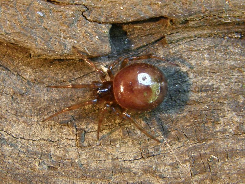
And here are the dimples, on another, very tiny S. bipunctata, I found this summer. (Maybe it's Brownie, as a baby.)


And thanks, all, for your help!

*Next Valentine's Day, I'm going to make my hearts this shape; long and skinny, with spikes.
.
Powered By Blogger